Neurogenesis - the Adult Brain also Forms New Neurons
How New Nerve cells are created in the Brain
Transcranial Pulse Stimulation Supports the Process of Neurogenesis
Until about 20 years ago, it was believed that all the nerve cells of the brain were already created at birth. But it has now been scientifically proven that the adult brain also forms new nerve cells – and does so throughout life. This process is called neurogenesis. In two regions in particular, the olfactory bulb and the hippocampus, new neurons are constantly being formed. To be sure, neurogenesis declines with age, as do many other bodily functions. But as research has shown, neurons are still being formed at an advanced age. This leads us to the regenerative capabilities of our body: almost all tissue, skin and bones as well as organs can be repaired to a certain extent by the body itself, because it is – together with the brain – capable of great regenerative efforts. We owe this to a large extent to stem cells. All these findings are incorporated into the principle of transcranial pulse stimulation.

Stem cells keep the human system running.
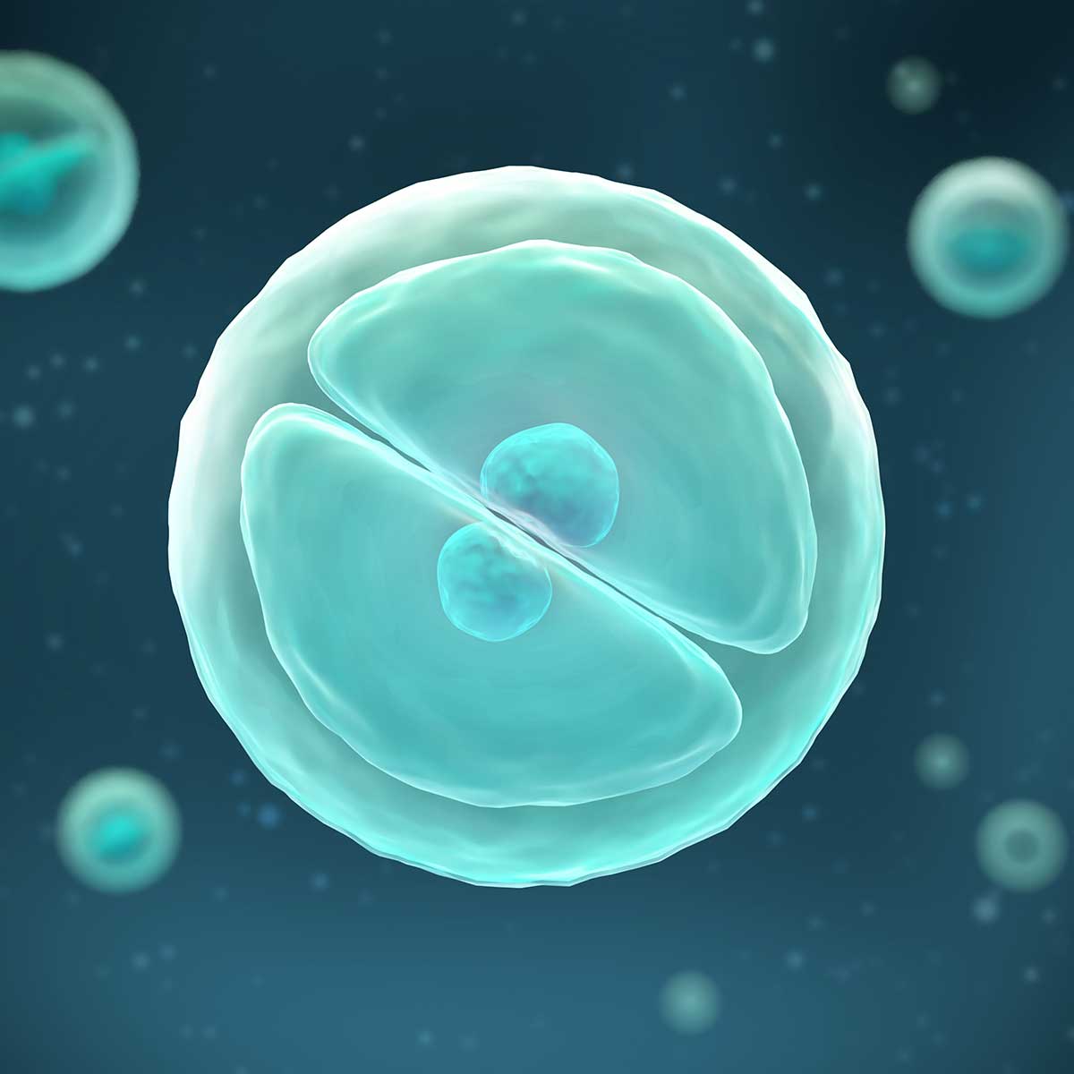
Stem cells are versatile cells and resemble a developing embryo. They have the ability to divide almost any number of times and transform into any type of cell. A good example of this are the hematopoietic stem cells in the spinal cord: they develop as needed into platelets, red blood cells and the broad spectrum of white blood cells.
The same is true for the neurons of the brain, although there are different types of neurons: We have neurons, which are the basic elements of our nervous system that use electrical and chemical signals to receive information, process it, and transmit it through axons (nerve fibers). Glial cells, in turn, form the cellular tissue that fills all but a small gap in the space between the brain’s neurons and blood vessels, and also form the myelin sheaths (myelin sheaths) around the nerve fibers.
The activity of stem cells and their transformation into nerve cells of all kinds begins in the human embryo. Already a few days after the fertilization of the egg cell, the development of the brain begins in the becoming organism. Every minute, about 250,000 nerve cells are formed and with birth, about 100 billion of these neurons (nerve cells) are waiting for information and impressions in the little head of the new citizen of the earth.
For a long time, the prevailing opinion was that this number of nerve cells must be sufficient for an entire human life, and many of us probably learned this in school. But at the end of the 1990s, this (doctrinal) opinion underwent a fundamental, almost radical revision: The formation of new nerve cells in adults was demonstrated for the first time by the Swede Thomas Björk-Eriksson from the University of Gothenburg and, after numerous further studies, led to a paradigm shift in medicine, albeit a somewhat bumpy one.
Neurogenesis: An orchestra whose number of instruments decreases, but keeps on playing.
Unfortunately, it is not the case that we are supplied throughout our lives with the enormous number of new nerve cells that we had in early childhood. As adults, we no longer have to process the enormous information and learning impulses that affect us in early childhood. Neural stem cells are present in all adult brain regions, but in many of them they are dormant or waiting for stimulation according to very specific laws of the control mechanisms of the human organism.
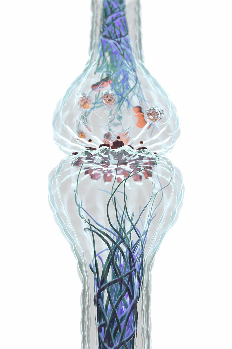
Adult neurogenesis”, i.e. the formation of new nerve cells, takes place mainly in the hippocampus, an area of the brain that is important for learning and memory processes. These new formations can also be detected in the so-called olfactory bulb (located below the frontal brain and used for olfactory perception). But in order for the newly formed nerve cells to become active, stimuli from outside are necessary: seeing, hearing, smelling, tasting, communicating, in simple terms: being interested, moving around, always learning, keeping the brain active are the prerequisites for ensuring that the freshly developing brain cells do not immediately perish again. Only through stimulation can they mature into functional neurons whose axons form synapses. Synapses are the connection points between cells and serve to transfer information and pass it on by means of electrical impulses. And the more synapses the neurons can form, the more information can be transmitted. In a healthy adult brain, around 100 trillion (!) synapses are formed, which can process around 1013 analog arithmetic operations per second!
When a dementia disease develops, the synapses first perish and an ever-increasing number of nerve cells die off. Newly emerging neurons, of course, are far from being able to compensate for this. But they can be offered support – also in the form of shock waves.

Stimulation by Shockwaves: How Nerve cells are Triggered into Action.
Shock waves have been used in medicine since the 1980s and are firmly established in the fields of orthopedics, urology, dermatology, etc. With transcranial pulse stimulation, shock waves have now extended their field of application to diseases of the central nervous system. Shock waves possess a whole range of special properties that make it possible to exert therapeutic effective forces on localized tissue areas and to initiate a process known as “mechanotransduction”: This refers to the transmission of mechanical stimuli (“mechano”) to tissues and cells and a biological response (“transduction”) to them in the form of regeneration, biostimulation, or cascading activation of specific bodily mechanisms.
But what are shock waves actually? Shock waves, also called sound waves, are first of all mechanical or acoustic waves. They are created by the ultra-short compression (i.e. compression – e.g. of gases or air – with an increase in pressure and reduction in volume) and subsequent relaxation of matter. Shock waves can pass through organic matter without much change, absorption, or damage. A shock wave has a singular pressure pulse that lasts only about one millisecond. Since a medium such as air or water is needed to generate a shock wave, shock waves used in medicine are generated in water. And it is precisely nerve cells that can be stimulated to activity or to emit action potentials with particularly weak shock wave pulses.
In transcranial pulse stimulation, these extremely low-frequency shock waves are transmitted non-invasively through the skullcap into the brain tissue via a hand-held applicator, and specific regions in the brain can be targeted and treated. In the process, metabolic processes at the synaptic switching points (the axons) of the nerve cells are activated and properly trained, i.e. stimulated. The extremely short sound pulses of transcranial pulse stimulation lead to short-term membrane changes in the brain cells. The concentration of transmitters and other biochemical substances is changed locally. The consequence is an activation of nerve cells and the development of compensatory networks, i.e. the formation of new synapses, which improve the diseased brain function. In addition, growth factors are released, which in turn influence the development of stem cells. In addition, there is an improvement in cerebral blood flow, as well as the formation of new vessels and nerve regeneration. Finally, the treatment can support the release of nitric oxide and the stimulation of the so-called BDNF, proteins from the group of neurotrophins that protect nerve cells and synapses.
To sum up, Transcranial Pulse Stimulation is simply a supportive and at the same time gentle tool for the human organism to regenerate itself. However, like all innovations in this world, it takes time for this knowledge to find its way into the general knowledge described at the beginning. It is to be hoped that this will not again take more than 20 years as with adult neurons.
Notice:
These explanations are deliberately kept highly simplified and do not go into further detail about the complex processes in the human brain and the many years of intensive research in this regard. This information does not replace the personal consultation with Prof. Dr. med. Musa Citak and/or his medical practice colleagues.

